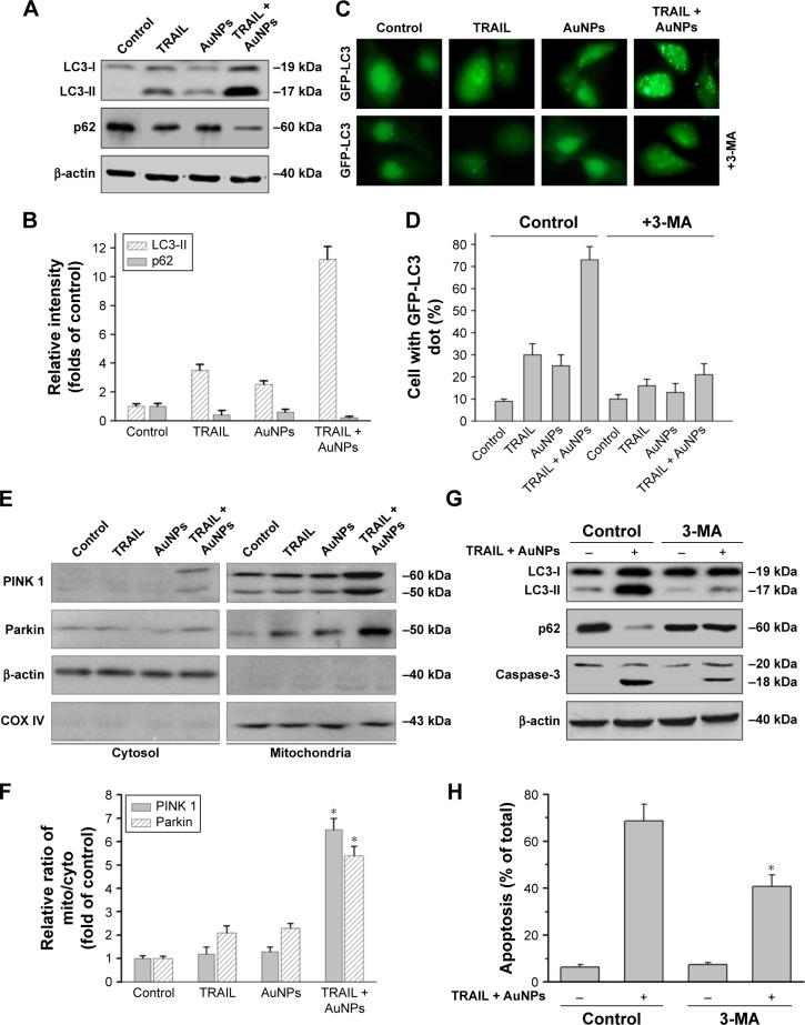
Figure 7.
TRAIL combined with AuNPs triggers autophagy.
Notes: (A, B) Calu-1 cells were exposed to TRAIL and AuNPs, alone and together, for 24 h, and then cell lysates were processed for immunoblotting analysis using antibodies against LC3 and p62. β-actin served as a loading control. Densitometric analysis was carried out to evaluate the relative levels of LC3-II and p62. (C, D) Formation of GFP-LC3 puncta induced by TRAIL combined with AuNPs. Cells were transiently transfected with the plasmid expressing GFP-LC3. At 48 h after transfection, cells were exposed to TRAIL and/or AuNPs in the absence or presence of 2 mM 3-MA, and GFP-LC3-labeled autophagic puncta formation was observed with a fluorescence microscope. Statistical analysis of the number of GFP-LC3 puncta per cell 24 h after treatment. Magnification ×200. (E, F) TRAIL combined with AuNPs induced mitophagy. Upon treatment with TRAIL and AuNPs, alone and together, the cytosolic and mitochondrial expression of PINK1 and Parkin was examined by immunoblotting analysis. Densitometric analysis was performed to estimate the relative intensity of PINK1 and Parkin. β-actin and COX IV were used as mito and cytoplasmic markers, respectively. *P<0.05, compared to TRAIL-treated groups. (G) Inhibition of autophagy decreased apoptosis induced by co-treatment with TRAIL and AuNPs. Cells were exposed to combination treatment with or without 3-MA. Total cell lysates were subjected to immunoblotting analysis. (H) Apoptosis was evaluated by flow cytometry using the sub-G1 assay. *P<0.05, compared to the TRAIL and AuNPs group.
Abbreviations: 3-MA, 3-methyladenine; AuNPs, gold nanoparticles; cyto, cytoplasm; mito, mitochondria; TRAIL, tumor necrosis factor-related apoptosis-inducing ligand.

