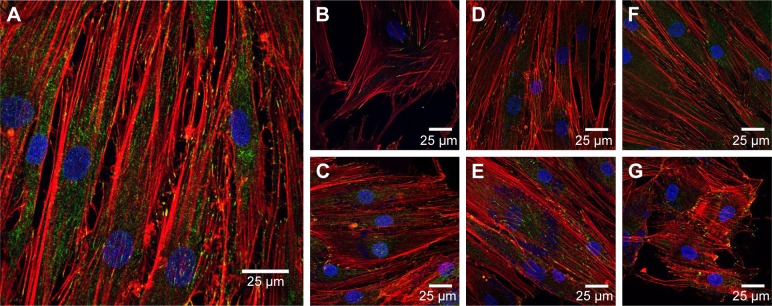
Figure 6.
Confocal microscopy of HDFs.
Notes: Vinculin focal adhesion points were stained with FITC–anti-vinculin (green), actin fibrils were stained with Alexa Fluor 568 phalloidin (red) and cell nuclei were stained with Hoechst 33258 (blue). (A) Control without NPs, (B) 25 μg/mL AuNSTs, (C) 500 μg/mL AuNSTs, (D) 25 μg/mL AuNFs, (E) 500 μg/mL AuNFs, (F) 25 μg/mL AuNSs and (G) 500 μg/mL AuNSs. The NPs also appear in blue.
Abbreviations: HDF, human dermal fibroblast; FITC, fluorescein isothiocyanate; NP, nanoparticle; AuNST, gold nanostar; AuNF, gold nanoflower; AuNS, gold nanosphere.

