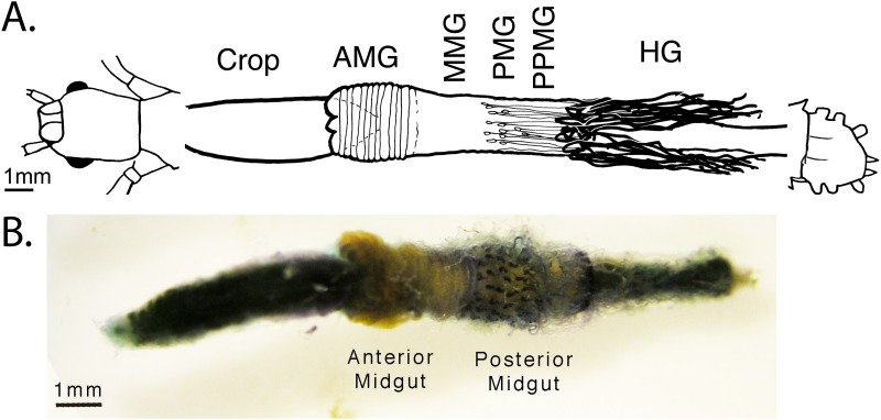
Fig 1. Digestive and excretory system of the Phasmatodea.
A) Schematic and B) dissection of the alimentary canal from Aretaon asperrimus, (Heteropterygidae) typical of other Phasmatodea [32]. The insect was vitally stained with New Methylene Blue N and dissected 6 days later. The appendices [violet] appear on the posterior midgut. The Malpighian tubules [colorless] originate at the midgut/hindgut junction, trailing over the posterior midgut before going towards the posterior end of the insect. The gut section between the two, the “post-posterior midgut,” was used for our midgut wall (MGWall) samples, excluding any tubules. Key: AMG = anterior midgut. HG = hindgut, MMG = middle midgut. PMG = posterior midgut. PPMG = post-posterior midgut. The schematic is reused with permission from this author’s previously published work [32].

