
Figure 4.
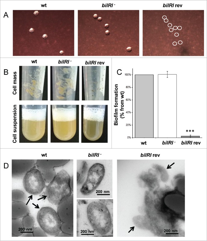
The outer membrane lipoprotein BilRI was not essential for the formation of typical A. actinomycetemcomitans rough-type colonies, biofilm or cell size and shape. BilRI overexpression-induced lysis of the outer membrane resulted in tiny colonies and significantly reduced biofilm amounts. A) On blood agar plates, the bilRI− mutant formed typical rough-type colonies, whereas the BilRI-overexpressing strain (bilRI rev) formed very tiny colonies (circled in white). B) Uniform cell suspensions could be produced similarly with the wild-type and bilRI− mutant strains following the method described by Karched et al.31 Because the BilRI-overexpressing strain bilRI rev grew slowly on agar plates, it was difficult to harvest a sufficient cell mass to obtain a dense cell suspension. C) The bilRI− mutant formed as much biofilm as the wild-type strain after 20-24 hours, as estimated through Crystal violet staining.32 The overexpression of BilRI almost completely eliminated the capacity of the strain (bilRI rev) to form biofilm (***:p = 0.0003, paired-samples T-test with Bonferroni corrections). D) Transmission electron microscopy revealed that the shape and size of the bilRI− mutant cells resembled those of wild-type cells. The overexpression of BilRI (bilRI rev) lysed the bacterial outer membrane, resulting in a smaller cell size. Arrows indicate the A. actinomycetemcomitans cells in images in which other structures, such as the filter disc, are visible.