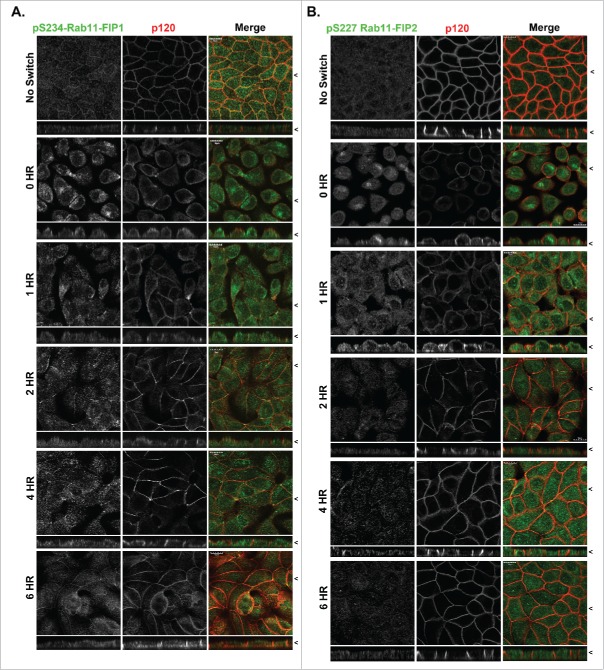
Figure 2.
Localization of p(S234)-Rab11-FIP1 is distinct from p(S227)-Rab11-FIP2 during re-polarization of MDCK cells. T23 MDCK cells labeled with either p(S234)-Rab11-FIP1 antibodies (A) or p(S227)-Rab11-FIP2 antibodies (B) along with antibodies against p120 in non-switched cells (NS) and then 0, 1, 2, 4, or 6 hours after re-addition of calcium. Cells were fixed with methanol. Merged dual color images are shown at the right. Localization of p(S234)-Rab11-FIP1 was punctate/vesicular as well as junctional with the greatest density in the apical region and was maintained throughout the calcium switch. Phosphorylation of p(S227)-Rab11-FIP2 was upregulated in calcium switch, however, p(S227)-Rab11-FIP2 labeling peaked at 2 hours after calcium switch and then declined. X/Z images are shown below X/Y images, < indicates where images were taken for X/Y and X/Z planes. All scale bars = 10 µm.

