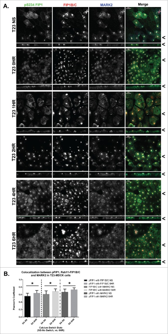
Figure 3.

MARK2 colocalizes with Rab11-FIP1B/C and p(S234)-Rab11-FIP1 throughout calcium switch. A. Non-switched T23 MDCK cells (NS) and cells at 0, 1, 2, 4, and 6 hours after calcium re-addition were fixed with 4% paraformaldehyde and immunostained using the p(S234)-FIP1 antibody, a Rab11-FIP1B/C antibody, and a MARK2 antibody. The MARK2 colocalized with both the p(S234)-FIP1 and FIP1B/C staining. Colocalization was observed between p(S234)-FIP1 and MARK2 at the lateral membrane 4 or more hours after calcium switch. X/Z images are shown below X/Y images, < indicates where images were taken for X/Y and X/Z planes. All scale bars = 10 µm. B. Colocalization analysis was conducted comparing p(S234)-FIP1 (pFIP1) with Rab11-FIP1B/C, pFIP1 with MARK2, and MARK2 with Rab11-FIP1B/C. All proteins colocalized together, and in each case, colocalization increased significantly through calcium switch (pFIP1 and FIP1B/C p = 0.024, FIP1B/C and MARK2 p = 0.026, pFIP1 and MARK2 p = 0.031, graphs plot mean Pearson's Coefficient, error bars represent standard deviation).
