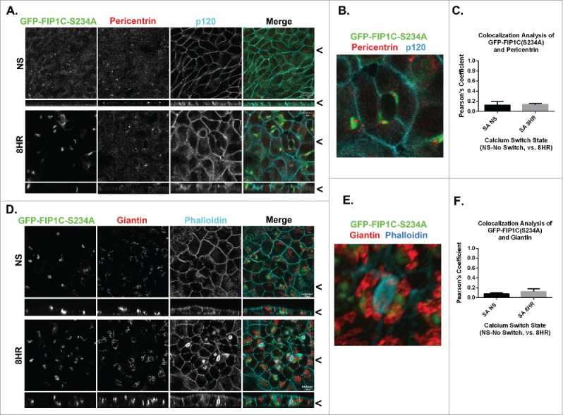
Figure 9.

Overexpression of GFP-Rab11-FIP1C(S234A) causes reorientation of the centrosome-based axis after 8-hour calcium switch. GFP-FIP1C(S234A) expressing cells, either non-switched (NS) or fixed at 6 hours following calcium re-addition were examined for localization of the centrosome marker Pericentrin (A-C) along with p120 staining or for the Golgi apparatus marker Giantin with phalloidin staining (G-I). A. Endogenous pericentrin in non-switched cells localized to a nidus below the apical membrane. However, 8 hours after calcium switch pericentrin relocalized to a point adjacent to lateral lumens in close proximity to GFP-FIP1C(S234A). B. In a still image taken from 3-dimensional reconstruction, the Pericentrin-staining centrosome is located directly adjacent to GFP-FIP1C(S234A) in association with a lateral lumen (Movie S7). C. No colocalization was seen between pericentrin and GFP-FIP1C(S234A). D. In non-switched cells, Giantin was located deep to the apical membrane. However, 8 hours after calcium switch, Giantin, labeling the Golgi apparatus, reoriented to face the lateral lumen surface. E. In a still image taken from 3-dimensional reconstruction, Giantin stain localizes adjacent to GFP-FIP1C(S234A) however, very little colocalization (F) is seen (Movie S8). X/Z images are shown below X/Y images, < indicates where images were taken for X/Y and X/Z planes. All scale bars = 10 µm. In C and F, graphs plot mean Pearson's Coefficient and error bars represent standard deviation.
