Figure 10.

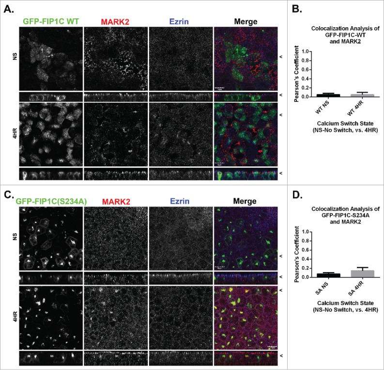
MARK2 localization is altered in FIP1C(S234A) overexpression lines. Non-switched GFP-FIP1C wildtype and GFP-FIP1C(S234A)-expressing MDCK cells or cells at 4 hours following calcium re-addition were fixed and immunostained for MARK2 and Ezrin. A and B. GFP-FIP1C wildtype did not colocalize with MARK2 in either non-switched cells or 4 hours after calcium switch. C. In non-switched GFP-FIP1C(S234A)-expressing cells, MARK2 did not localize with GFP-FIP1C(S234A). However, in switched cells at 4 hours after re-addition of calcium, a point before lateral lumens are formed, MARK2 colocalization with GFP-FIP1C(S234A) was observed. D. Colocalization of MARK2 and GFP-FIP1C(S234A) overall was weak. However, there was a statistically significant increase from NS to 4 HR. X/Z images are shown below X/Y images, < indicates where images were taken for X/Y and X/Z planes. All scale bars = 10 µm. In B and D, graphs plot mean Pearson's Coefficient and error bars represent standard deviation.
