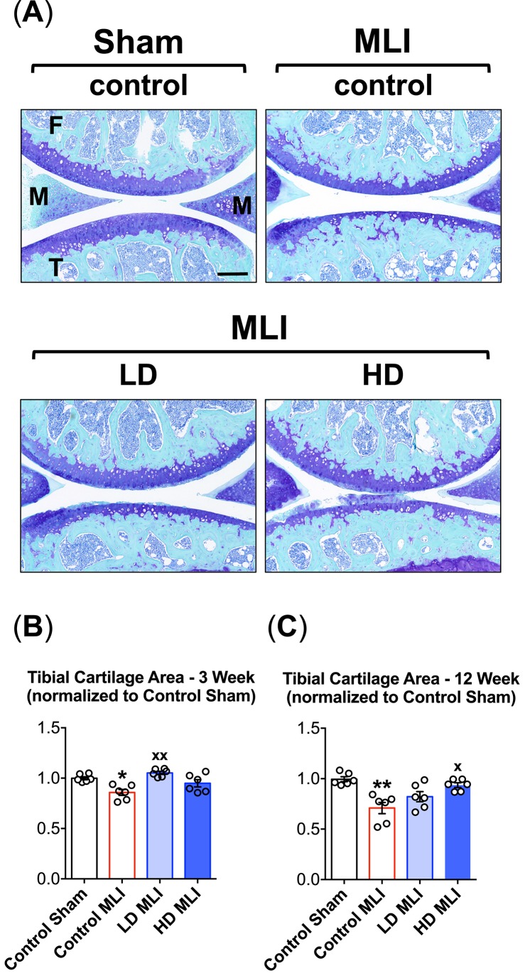
Fig 2. Cartilage loss following MLI in hCol1-fed mice is reduced.
Panel (A) presents an array of representative 40x Toluidine Blue/Fast Green stained sagittal sections from the medial compartment of sham and MLI joints 12 weeks post-injury under various treatment conditions (control = vehicle, LD = 3.8mg hCol1/day, HD = 38mg hCol1/day). Joint structures are labeled (F = femur, M = meniscus, T = tibia) and the black scale bar depicts 100μm. Total tibial cartilage area was determined in these representative sections as well as a series of similarly stained serial sections from all experimental joints at both 3 weeks (B) and 12 weeks (C) post-MLI using an automated approach (Visiopharm System). Symbols (○) represent the average tibial cartilage area of 3 sections/joint. Bars represent the average tibial cartilage area for each experimental group (± SEM, N = 6). Significant differences between experimental groups were identified via one-way ANOVA with a Tukey’s multiple comparisons post-test (*p<0.05, **p<0.01 compared to Control Sham; xp<0.05, xxp<0.01 compared to Control MLI).

