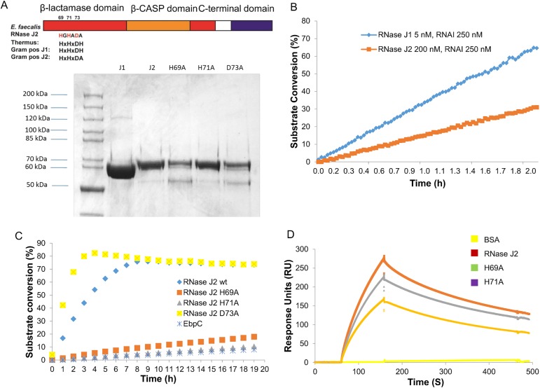
Fig 2. E. faecalis RNase J1 and J2 are active exonucleases.
(A) Diagram depicting the metallo-β-lactamase (red), β-CASP (orange) and dimerization domains. Residues that may be involved in zinc coordination were highlighted. Purified N-His6 recombinant E. faecalis RNase J1, J2 and three point mutations (H69A; H71A; D73A) of J2 were analyzed by SDS-PAGE. (B) and (C) Exonuclease activities of RNase J1, J2 and three J2 mutants were determined by RT-FeDEx analysis. Recombinant EbpC protein served as a negative control for the assay. (D) Binding of E. faecalis RNase J2 and two mutants (H69A and H71A) (all represented at 100 nM) to immobilized E. faecalis RNase J1 on a CM5 sensor surface is shown by the SPR sensorgrams.

