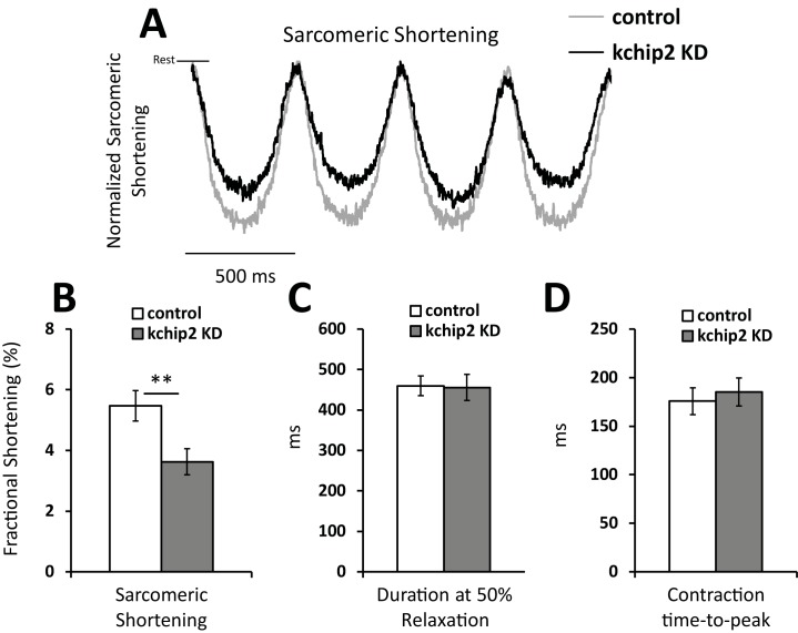
Fig 2. KChIP2 knock down reduces myocyte contractility.
(A) Representative tracing of the change in distance between two consecutive sarcomeres during contraction. Cells were paced at a 500 ms cycle length for Ad.GFP (n = 26) or Ad.KChIP KD (n = 28) treated myocytes. Tracings were normalized to control cells. Summary data for (B) fractional shortening, (C) contraction duration at 50% amplitude, and (D) the time to peak contraction. Data presented as mean ± SEM; *P < 0.05, **P < 0.01; two-tailed Student’s t-test.

