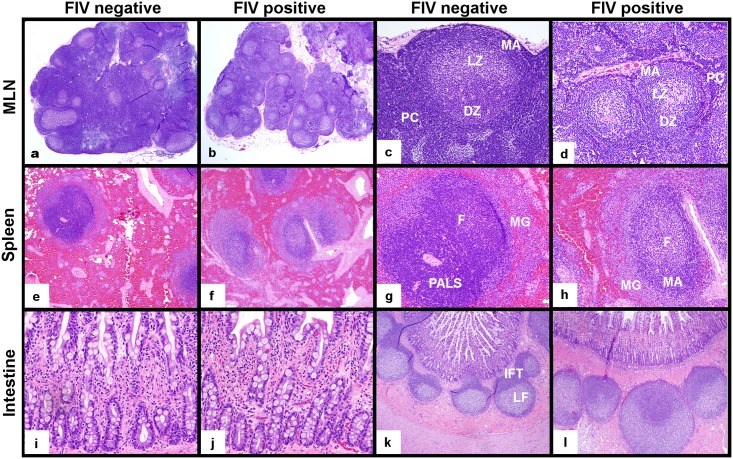
Fig 5. Histologic examination of MLN, spleen and intestine from FIV-infected progressor cats in the late asymptomatic phase and uninfected cats.
MLNs from FIV-infected progressor cats were smaller in size and characterized by paracortical atrophy relative to those from uninfected cats (a,b, 20x). MLN follicles from FIV-infected cats revealed active germinal centers with less densely populated dark (DZ) and light zones (LZ), a prominent mantle zone (MA) and a thin, sparsely populated paracortical rim (PC) relative to uninfected cats (c,d, 100x). Spleens of infected progressor cats showed less densely populated white pulp that exhibited indistinct periarteriolar lymphoid sheaths (PALS), prominent active germinal center follicles (F) with expanded mantle (MA) and marginal zones (MG), relative to uninfected cats (e,f 40x, g,h 100x). Notable differences in intestinal mucosal architecture and leukocyte density were not observed between uninfected and FIV-infected cats (200x, i,j). Intestinal Peyer’s patches (lymphoid follicles) of uninfected and infected cats both contained active germinal centers; however, interfollicular T-cell zones (IFT) were better demarcated in intestinal biopsies from uninfected cats relative to infected cats (k,l, 40x).

