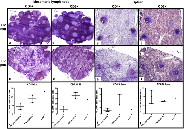
Fig 6. Determination of CD4+ and CD8+ leukocyte frequency in MLN and spleen from FIV-infected and uninfected cats.
A severe CD4+ depletion is observed in the paracortex and medullary cords of MLN and in periarteriolar lymphoid sheaths of the spleen of FIV-infected progressor cats relative to uninfected cats (a,b,g,h), consistent with the flow cytometry data (c,i). A notable CD8+ depletion is also evident in the paracortex of MLNs from FIV-infected progressor cats relative to uninfected cats (d,e), and supported by flow cytometric analysis (f). IHC analysis for CD8+ T cell distribution demonstrates diffuse staining of the red pulp in both infected and uninfected cats (j,k) whereas splenic CD8+ T cell frequencies are variable among cats by flow cytometry (l). For flow cytometry data, each point represents one cat, the horizontal bar represents mean, and the whiskers represents the range. The LTNP cat’s flow cytometry value is a single triangle.

