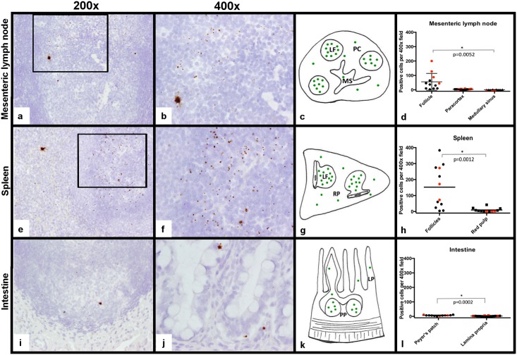
Fig 8. Detection of FIV RNA in the MLN, spleen and intestines.
FIV RNA localization in tissues was measured by in situ hybridization using RNAscope and represented by brown chromogen dots. Viral RNA was concentrated in lymphoid follicles (LF) within the MLN (a, 200x magnification). At higher magnification (400x), the viral RNA signal was predominantly represented as a single dot, but rare cells were distended with brown chromogen that extended into stellate cell processes consistent with dendritic cell morphology (b). FIV RNA was less frequent in the paracortex (PC) and medullary sinus (MS) regions (c, d). Similarly, FIV RNA was concentrated in the lymphoid follicles of the spleen, and was less frequent in the red pulp (RP, e-h). Within intestinal tissue, FIV RNA was again most concentrated in lymphoid follicles of the Peyer’s patches (PP, i-l) and less frequently detected in the lamina propria (LP) of villi (j, 400x). Red dots on the graphs represent the LTNP cat.

