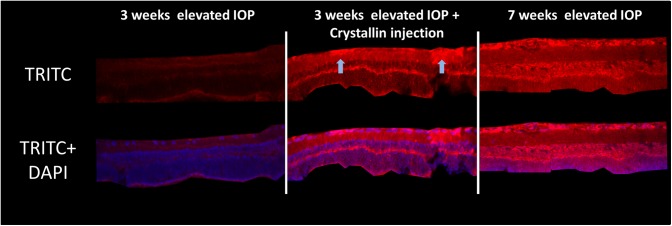
Fig 4. β-crystallin B2 staining in retinal cross-sections.
IHC staining could validate the results from mass-spectrometric featured proteomics analyses. After 3 weeks of IOP elevation, almost no β-crystallin B2 could be detected in the retina cross section (n = 3), while a strong increase of protein level could be verified after 7 weeks of elevated IOP (n = 3). Injection of β-crystallin B2 seems to change the protein level in retinal cells after 3 weeks of IOP elevation considerably due to cellular uptake of the crystallin, predominantly by the RGC layer (n = 4).

