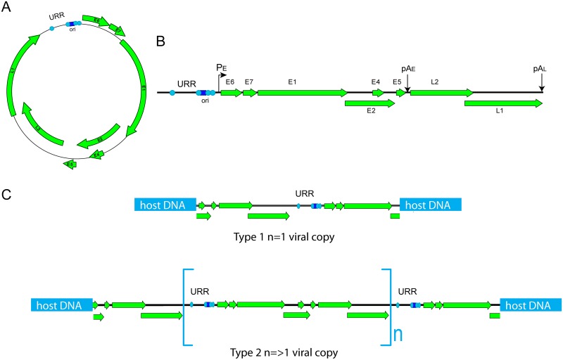
Fig 1. Types of HPV integration.
A. Circular HPV genome. B. Linear HPV genome. URR (upstream regulatory region), PE (early promoter), and pAE and pAL (early and late polyadenylation sites) are indicated. The light blue circles in the URR represent E2 binding sites, and the dark blue square is the E1 binding site in the origin of replication (ori). C. In Type 1 integration, a single viral genome is integrated into the host DNA. In Type 2 integration, multiple genomes are integrated in tandem in a head-to-tail orientation. This often is accompanied by focal rearrangement and amplification of flanking cellular sequences.

