Figure 5.

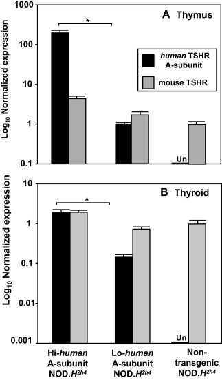
Expression in thymic and thyroid tissue, measured by quantitative polymerase chain reaction (qPCR), of the human thyrotrophin receptor (TSHR) A‐subunit transgene and the endogenous mouse TSHR in Hi‐expressor non‐obese diabetic (NOD).H2h4 offspring, Lo‐expressor NOD.H2h4 offspring and non‐transgenic NOD.H2h4 mice. (a) Thymus; data are shown as log10 relative expression [mean + standard deviation (s.d.) of triplicates] normalized to mouse β‐actin for the human TSHR A‐subunit or the mouse TSHR. (b) Thyroid; data are shown as log10 relative expression (mean + s.d. of triplicates) normalized to mouse β‐actin and glyceraldehyde 3‐phosphate dehydrogenase (GADPH) for the human TSHR A‐subunit or the mouse TSHR. Expression of the human TSHR A‐subunit was undetectable (Un) in both thymus and thyroid of non‐transgenic NOD.H2h4 mice. The thymic data are representative of three comparable experiments. A similar (but separate) study confirmed the thymic expression values for Lo‐expressor transgenics versus non‐transgenic NOD.H2h4 mice 46. Thyroidal mRNA expression levels of the Hi and Lo‐expressor transgenes are similar to previous observations of immunohistochemistry and protein extraction 15. Significantly different human TSHR A‐subunit expression in the Hi‐ versus the Lo‐expressor: *P = 0.009; ^P = 0·001 (t‐tests).
