
Figure 4. Irf8/β-catenin self-renewal progenitor network.
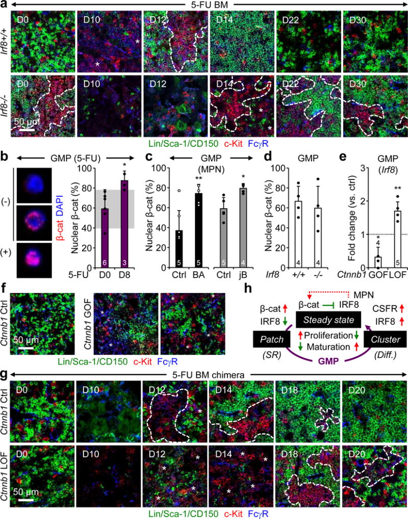
a, Representative IF staining of GMPs (purple) in 5-FU-treated Irf8+/+ and Irf8−/− BM. b–d, Nuclear β-catenin in (b) GMPs from d8 5-FU-treated WT mice with representative negative (−) and positive (+) staining shown for d0 GMPs, (c) GMPs from BA, jB and respective age-matched Ctrl mice, and (d) GMPs from Irf8+/+ and Irf8−/− mice. Results are expressed as percent positive cells. e, Irf8 expression in GMPs from Ctnnb1 gain-of-function (GOF) and loss-of-function (LOF) mice. f, Representative IF staining of GMPs (purple) in Ctnnb1 Ctrl and GOF BM. g, Representative IF staining of GMPs (purple) in 5-FU-treated Ctnnb1 Ctrl and LOF BM. h, Model of the molecular network controlling pGMP self-renewal (SR) and cGMP differentiation (diff.). Red indicates activation and green suppression, with dotted line highlighting re-enforcing pathways in MPNs. Stars indicate pGMPs and dotted lines cGMPs. Results are expressed as mean ± S.D. (grey bars, reference range); *p ≤ 0.05; **p ≤ 0.01, ***p ≤ 0.001.