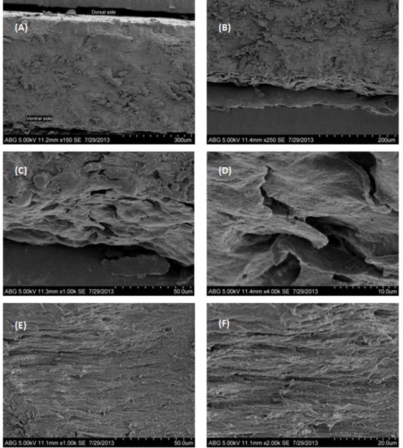
Figure 4.

SEM images of the cross-section of nail showing (A) three layers of nail plate (150×); (B) intermediate and ventral layer (250×); (C) ventral layer (1000×); (D) ventral layer cells (4000×); (E) fiber orientation in the intermediate layer (1000×) and (F) fiber orientation in the intermediate layer (2000×).
