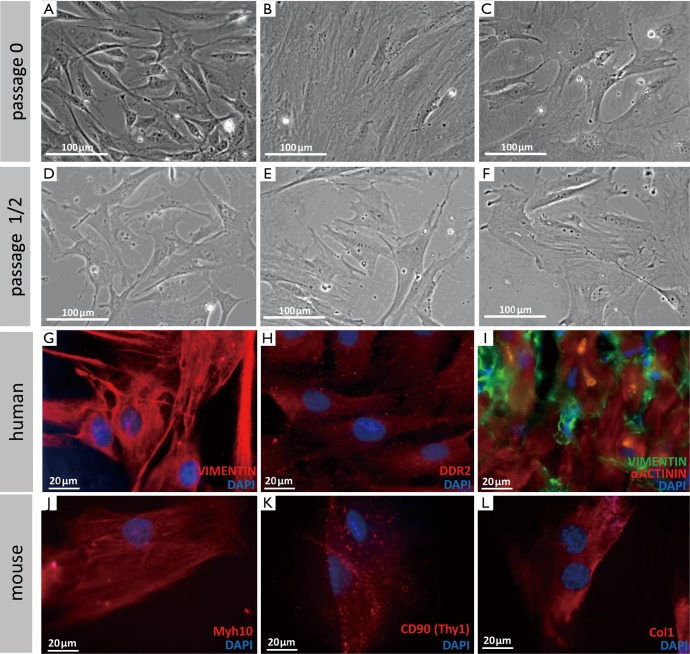
Figure 3.
Morphology and markers of different types of fibroblasts. (A-F) Morphology of different types of fibroblasts grown in cell culture for different times (phase contrast). (A,D) Mouse tailtip fibroblasts. (B,E) Human adipose tissue-derived fibroblasts. (C,F) Human cardiac fibroblasts. (G-L) Immunofluorescent staining with different widely used markers of human and mouse fibroblasts. Vimentin (G) and DDR2 (H) were used to stain cultured human cardiac fibroblasts. (I) Vimentin staining combined with α-Actinin (cardiomyocyte) staining on a cryosection of a human right ventricular biopsy. Myh10 (J), CD90 (K), and Col1 (L) were used to stain cultured murine tail tip fibroblasts.

