Fig. 1.
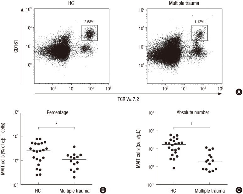
Reduced circulating MAIT cell numbers in the peripheral blood of multiple trauma patients. Freshly isolated PBMC from 22 HCs and 14 patients with multiple trauma were stained with APC-Alexa Fluor 750-conjugated anti-CD3, FITC-conjugated anti-TCR γδ, APC-conjugated anti-TCR Vα7.2 and PE-Cy5-conjugated anti-CD161 mAbs and then analyzed by flow cytometry. Percentages of MAIT cells were calculated within a αβ T cell gate. (A) Representative MAIT cell percentages as determined by flow cytometry. (B) MAIT cell percentages among peripheral blood αβ T cells. (C) Absolute MAIT cell numbers (per microliter of blood). Symbols (●) represent individual subjects; horizontal bars show the median.
MAIT = mucosal-associated invariant T, PBMC = peripheral blood mononuclear cell, HCs = healthy controls, APC = allophycocyanin, FITC = fluorescein isothiocyanate, TCR = T cell receptor, PE = phycoerythrin, mAbs = monoclonal antibodies, ANCOVA = analysis of covariance.
*P < 0.01, †P < 0.001 by ANCOVA test.

