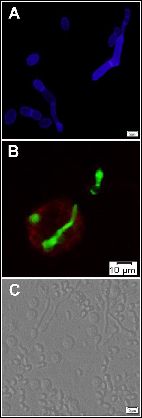
Figure 2.

The interaction of T. asahii with host immune cells. (A) Blastoconidia and pseudohyphae morphotypes of T. asahii stained with Calcofluor White. (B) Phagocytosis of T. asahii by human macrophages. Fungal structures and macrophages were stained with fluorescein isothiocyanate (FITC) and phycoerythrin (PE)-conjugated anti-CD14 antibody, respectively. Trichosporon cells were incubated overnight at 4°C with FITC (Sigma-Aldrich) at a concentration of 0.1 mg/mL in 0.1M Na2CO3 buffer. (C) Bright-field microscopy visualization of human peripheral blood mononuclear cells aggregated around the different morphotypes of T. asahii.
