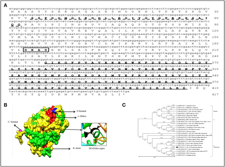
Figure 1.
Sequence analysis of PgeIF4A gene and protein. (A) Schematic Representation of three motifs including RNA helicase/DEAD box Rec-Q-motif (34–62, indicated as dotted line), DEAD box domain (183–186, in box), and RNA-helicase C-terminal (246–407) in PgeIF4A protein. (B) Predicted three dimensional structure of PgeIF4A protein showing various domains in different colors, blue represented N-terminal region, magenta for C-terminal, red for α-Helices and green for β-sheets. DEAD box region highlighted in red color circle which harbor Asp-183 in β-5, Glu-184 in linked region and Ala-185, Asp-186 presented in α-9 regions. (C) Phylogenetic tree constructed based on deduced amino acid sequences of various eIF4A from closely related species. Protein names, accession numbers, and species names were indicated at each branch. The phylogenetic tree generated using Neighbor Joining (NJ) method and viewed using MEGA4 software.

