Fig. 7.
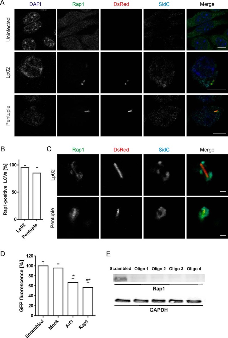
The small GTPase Rap1 localizes to LCVs in macrophages and promotes intracellular growth of L. pneumophila. A, RAW 264.7 macrophages were left uninfected or were infected (MOI 10, 2 h) with DsRed-producing L. pneumophila Lp02 or Δpentuple (pSW001), followed by labeling with anti-Rap1 and anti-SidC antibodies as well as DAPI. Bars, 5 μm. B, For quantification of (A), at least 50 infected macrophages were scored each in two independent experiments (means and S.D. are shown). C, RAW 264.7 macrophages were infected as in (A), and intact LCVs were isolated using the two-step immuno-affinity protocol. Isolated LCVs were immuno-labeled as in (A). Bars, 1 μm. D, Human A549 lung epithelial cells were treated for 48 h with siRNAs against Rap1, Arf1, scrambled siRNA or transfection reagent only (“mock”) and infected (MOI 10, 24 h) with GFP-producing L. pneumophila Lp02 (pNT28). Bacterial growth was determined by measuring GFP fluorescence in a plate reader. Data represent mean and standard deviation from three independent experiments (Student's t test: *p < 0.05, **p < 0.01) shown for one oligonucleotide out of four tested. E, A549 cells were treated with 4 different siRNAs (Oligo 1–4) against Rap1 for 48 h. The depletion efficiency was assessed by Western blot using a mouse monoclonal antibody against Rap1 (21 kDa). GAPDH (38 kDa) served as a loading control, and Qiagen AllStars oligonucleotides (“scrambled”) were used as a negative control.

