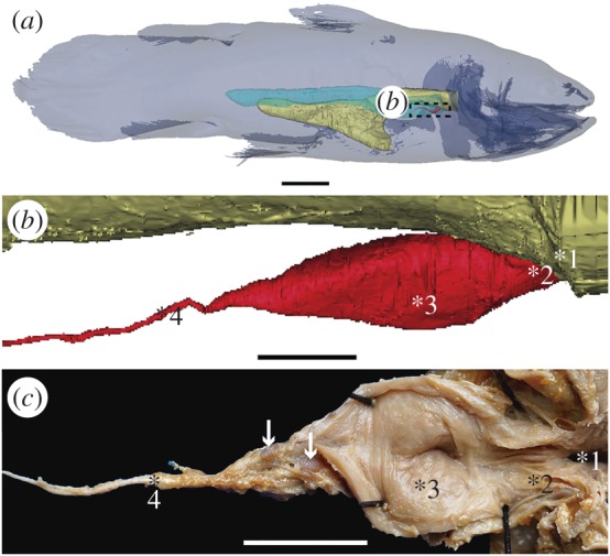
Figure 1.

The vestigial lung of the extant coelacanth Latimeria chalumnae. (a) Three-dimensional reconstruction of the adult specimen CCC 22 (130 cm TL) in right lateral view. (b) Details of the three-dimensional reconstruction of the lung of the adult specimen CCC 28, corresponding to the boxed area in (a). (c) Partial dissection of the lung of the adult specimen CCC 3, exhibiting its lumen in the ventral view. Yellow, oesophagus and stomach; red, vestigial lung; blue, fatty organ. White arrows point to two hard but flexible plates. 1, 2, 3, 4 indicate the four successive areas of the vestigial lung. Scale bars, 10 cm (a); 1 cm (b); 0.5 cm (c).
