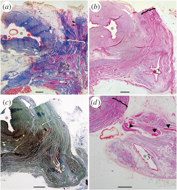
Figure 2.

Histological thin sections of the anterior part of the vestigial lung, from adult specimen CCC 5. Oesophagus at the top left (asterisks indicate the lumen) and lung in the bottom right (numbered asterisks localize the lumen relative to the lung in figure 1b,c). (a) Vestigial lung at the level of its origin (asterisk 2 of figure 1) with disorganized muscle bundles (brackets) surrounding this organ. (b,c) Anterior portion (two successive sections) of the lung still in close proximity with the oesophagus, showing invaginations in the lung walls (asterisk 2 of figure 1). Muscle bundles distributed in an organized network (brackets). (d) Vestigial lung completely dissociated from the oesophagus (asterisk 3 of figure 1), presenting the clearly reduced invaginations of the lung walls. Arrowheads indicating the hard, but flexible, plates. Scales bar 0.1 cm (a–d). (a,b,d, azocarmin; c, cajal colorations).
