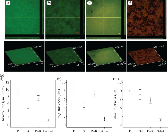
Figure 4.
Sensitivity of QSI-treated P. aeruginosa biofilms to kanamycin. Confocal scanning laser microscopy (CLSM) photomicrographs of PAO1 biofilms grown in the presence or absence of QSI supernatant (5%). Three days later, the biofilm was exposed to 100 µg ml−1 kanamycin for 24 h. (a) No QSI supernatant or kanamycin; (b) QSI supernatant (5%); (c) 100 µg ml−1 kanamycin; (d) QSI supernatant (5%) plus 100 µg ml−1 kanamycin. Live cells were stained green and dead cells were stained red using a LIVE/DEAD BacLight Bacterial Viability Kit. (e) COMSTAT quantification of (i) bio-volume, (ii) average thickness, and (iii) maximum thickness show that PAO1 forms thinner biofilms after being treated with kanamycin and QSI supernatant extract.

