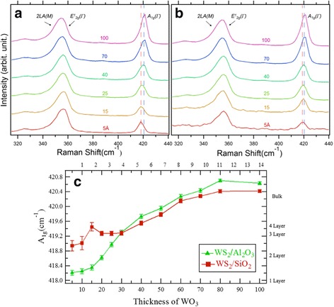
Fig. 3.

a Raman spectra from 0.5-nm- to 10-nm-thick WS2 grown on Al2O3 and b SiO2 substrate as a function of WO3 thickness (d WO3) (514.5 nm laser excitation, 300 K). The peaks at ~355 and ~420 cm−1 correspond to overlap of 2LA(M) and E1 2g(Γ) peaks and A1g(Γ) peak, respectively. Blue and red dashed lines indicate the A1g(Γ) peak position of the thick sample (d WO3 = 10 nm) and thin sample (d WO3 = 0.5 nm), respectively. Intensities are normalized with the A1g peaks. c Thickness dependence of the A1g(Γ) peak position
