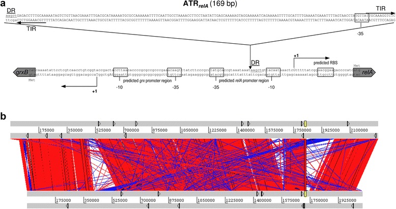
Fig. 7.

Genomic distribution of ATRs and the relA locus in N. meningitidis. a The intergenic region between grxB and relA. The integration site of a copy of an ATR repeat element upstream of relA (ATRrelA) in strain α522 is indicated with respect to the MC58 locus. The transcriptional start sites as determined by 5’-RACE in both strains are indicated along with the deduced −35 and −10 boxes and the computationally predicted promoter regions using PPP [117]. DR: direct repeat. b Alignment of both the MC58 (upper lane) and α522 (lower lane) genomes as visualized with the Artemis comparison tool based on a BLASTN comparison. The linearized MC58 and α522 genomes are shown in the upper and lower panel as gray bars, and regions syntenic in both genomes are connected via red and inverted regions via blue lines, respectively. The location of ATRs is indicated by small arrows in each genome, and the relA region is highlighted in yellow
