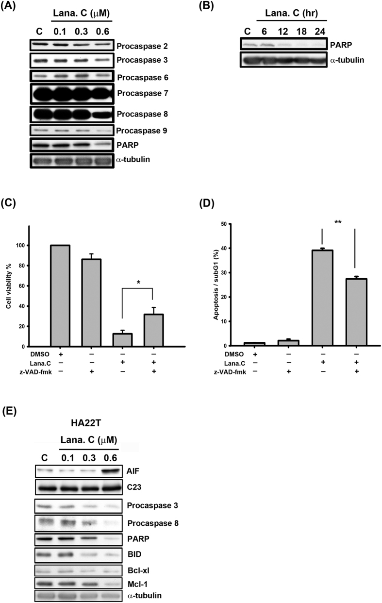
Figure 3. Lanatoside C induced apoptosis in HCC cells.
(A) Hep3B cells were treated with the indicated concentrations of lanatoside C (0.1–0.6 μM) for 18 h and detected of procaspase-2, -3, -6, -7, -8, -9 and PARP protein expressions by using Western blot analysis. (B) Hep3B cells were treated with lanatoside C (0.6 μM) for the indicated time (6–24 h), and cells were harvested from total lysates for detection of PARP protein expressions by using Western blot analysis. Data are representative of three independent experiments. (C and D) Hep3B cells were incubated in 0.6 μM lanatoside C with or without 100 μM z-VAD-fmk for 24 h. (C) The cell viability was determined by using MTT assay as described in methods. Data are repeated at least three independent determinations. *P < 0.05. (D) The apoptotic cells were stained with PI and analyzed by flow cytometry as described in methods. Data are repeated at least three independent determinations. **P < 0.01. (E) HA22T cells were treated with lanatoside C for 18 h to detect the expressions of caspases and mitochondrial proteins.

