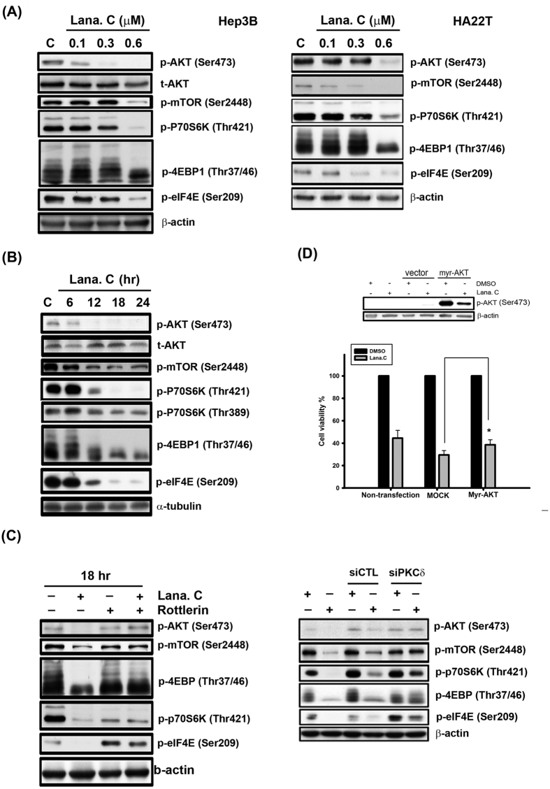
Figure 5. Effect of lanatoside C on AKT/mTOR pathway.
(A) Hep3B and HA22T cells were treated with a range of lanatoside C (0.1–0.6 μM) for 18 h. (B) Hep3B cells were treated lanatoside C (0.6 μM) for indicated time (6–24 hr) and then cells were harvested from total lysates for observation of AKT/mTOR and their downstream signaling protein expressions by using Western blot analysis. (C) Hep3B cells were incubated with Lanatoside C (0.6 μM), rottlerin (5 μM) or PKCδ siRNA, or combination treatment for 18 h and then cells were harvested from total lysates for detection of indicated protein expressions by using Western blot analysis. (D) Hep3B cells were transfected with empty vector (MOCK) or Myr-AKT for 6 h and re-serum overnight, followed by treatment with or without lanatoside C (0.6 μM) for 18 h. Cells were harvested from total lysates for detection of phospho-AKT Ser473 protein expressions by using Western blot analysis. Cell viability was measured by MTT assay. Data are expressed as means ± SEM of three independent determinations. *P < 0.01, Myr-AKT-overexpressed cells versus MOCK-transfected cells.

