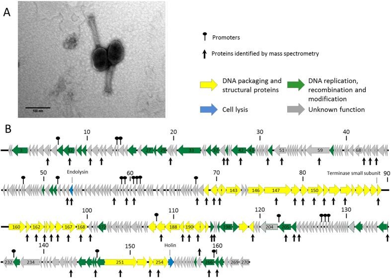
Figure 1. Morphology and genome overview of MP1.
(A) Electron micrographs of Morganella-infecting phage MP1 negatively stained with 2% uranyl acetate. (B) Genome map with predicted 271 CDSs numbered and coloured (yellow, green, blue and gray) according to their predicted function. Protein identified by mass spectrometry are pointed with vertical arrows. Above genome, the nucleotide positions in kb are given.

