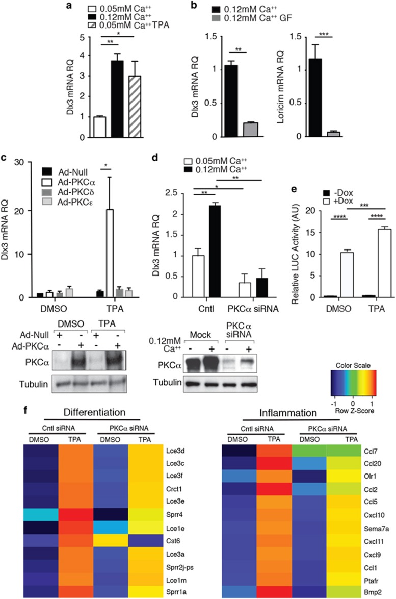
Figure 1.
PKCα triggers DLX3 expression in keratinocytes. (a) DLX3 expression in primary keratinocytes maintained in proliferative (0.05 mM Ca2+) and differentiating media (0.12 mM Ca2+) or treated with TPA for 24 h. (b) DLX3 and Loricrin expression in primary keratinocytes maintained in 0.12 mM Ca2+ media +/− GF109203X at 24 h. (c) Relative expression level of DLX3 in primary keratinocytes transduced with Adeno-Null or Adeno-PKCα, δ or ɛ viruses and treated with TPA for 1 h in 0.05 mM Ca2+ media. Bottom panel, western blot performed with protein extracts from cells transduced with Ad-Null and Adeno-PKCα, with or without TPA treatment. (d) DLX3 expression in primary keratinocytes transfected with PKCα siRNA at 48 h in 0.05 mM Ca2+ or 0.12 mM Ca2+ media. Bottom panel, western blot performed with protein extracts from cells transduced with PKCα siRNA. (e) Luciferase reporter assays were performed in PAM212-DLX3 tet-on cells transfected with a Firefly luciferase reporter construct containing the concatemerized canonical DLX3 binding. Cells were grown with or without Doxycyclin for 24 h to induce DLX3 expression. Significantly higher reporter activity was observed in cells treated with Dox after 6 h of TPA treatment. For all panels, *P<0.05, **P<0.01, ***P<0.001, ****P<0.0001 and the results are mean ± SD of three experiments. (f) Heatmaps of differentially expressed mRNAs in PKCα versus Scramble siRNA treated keratinocytes after TPA or DMSO (control) stimulus for differentiation (left) or inflammation (right) clusters. Expression values are colored based on their z-score after normalization across treatments

