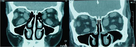
Figure 6. Coronal computed tomography of a patient with thyroid-associated ophthalmopathy (A). Coronal computed tomography images from the same patient after orbital decompression surgery (B). Postoperative images show the absence of the medial orbital wall and thinning of the cortical bone in the lateral wall.

