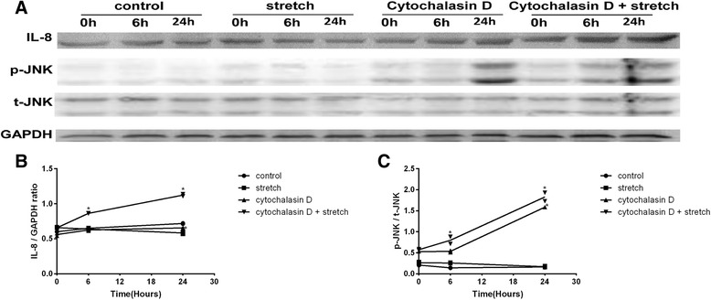
Fig. 2.

IL-8 expression and JNK phosphorylation levels in RPE cells. After cyclic stretch and Cytochalasin D exposure, cell lysates were prepared, and Western blotting analysis was performed on the indicated proteins. The IL-8 and JNK bands were scanned, and band densities were calculated using ImageJ software. The values are the band-density ratios compared with the baseline. The stimulation of the RPE cells with cyclic stretch alone did not induce a significant increase in IL-8 expression and JNK phosphorylation levels, which were similar to those of the control groups. After pre-treatment with Cytochalasin D alone, IL-8 expression and JNK phosphorylation levels were not significantly different at 6 h but were significantly increased at 24 h. After the pre-incubation of the RPE cells with Cytochalasin D followed by exposure to cyclic stretch, IL-8 expression and JNK phosphorylation levels increased at 6 h and 24 h (a-c). *P<0.01 versus baseline, n = 3 experiments
