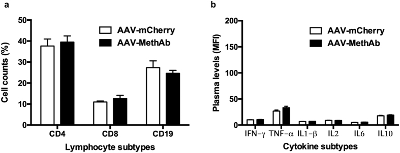
Figure 4. Lymphocyte counts and plasma cytokine levels in AAV-MethAb injected mice vs. those in AAV-mCherry injected mice.
Mice were treated with AAV-MethAb (2.5 × 10¹⁰ VGC/animal; n = 8, i.p.) or AAV-mCherry (2.5 × 10¹⁰ VGC/animal; n = 6, i.p.). At five weeks post-infection, peripheral blood was collected for the measurement of lymphocyte counts and plasma cytokine levels. (a) Three main types of immune cells, including CD4+ T lymphocytes, CD8+ T lymphocytes, and CD19+ B lymphocytes, were counted by flow cytometry analysis. Data were expressed as mean percentage of total cell counts (lymphocytes + monocytes) ± SEM. (b) The plasma levels of six representative cytokines, including IFN- γ, TNF-α, IL1-β, IL2, IL6, and IL10, were measured by Multiplex Immunoassay using a flow cytometry-based platform (Luminex 200). Data were expressed as mean fluorescence intensity (MFI) ± SEM. No significant differences for each measured lymphocytes or cytokines were found between AAV-MethAb and AAV-mCherry injected groups.

