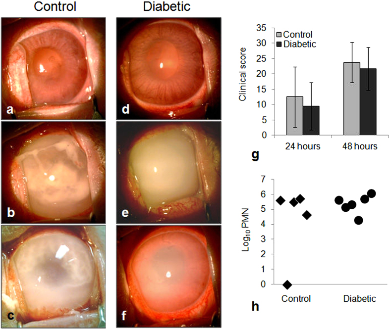
Figure 2.
(a–f) Eyes of control (a–c) and diabetic (d–f) rabbits, 48 hours after infection. (g) Clinical scores (determined by slit lamp examination) of control and diabetic rabbits; p ≥ 0.440. (h) Quantification of PMNs (determined by myeloperoxidase activity) in vitreous humor of both control and diabetic rabbits 48 hours after infection; p = 0.394. Error bars in (g) represent standard deviation.

