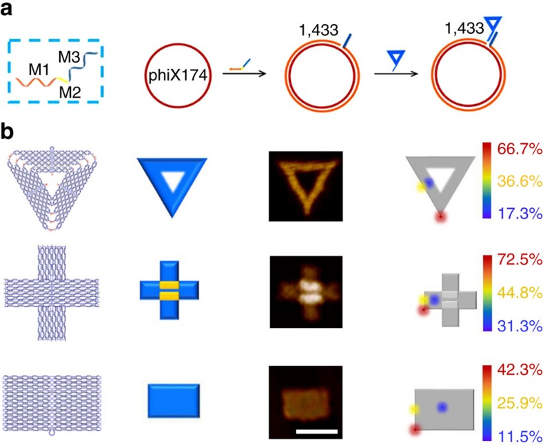
Figure 2. Origami shape IDs for ‘multi-colour' nanomechanical imaging.
(a) Schematic showing of the three-block ‘mediator' DNA strand (M-strand) in labelling of phiX 174 with shape IDs. Left, M-strand is shown with three function blocks. M1 block (orange-coloured region) for site-specific hybridization and extension with the template, M2 spacer block (yellow-coloured region) and a M3 block (blue-coloured region) for capturing shape IDs. Right, the phiX 174 template is first annealed with the M-strand on the site 1,433, then M1 block serves as allele-specific primer to initiating DNA extension in the presence of polymerase, which turns the ssDNA template into dsDNA that is more visible under AFM imaging. M3 block is complementary to a short strand M3′ on each corresponding shape ID. (b) Schematic and AFM images with the labeling efficiency of individual shape IDs. Left to right: design (column 1), schematic (column 2) and AFM images (column 3) of triangular-, cross- and rectangular- shaped DNA origami IDs. Scale bar, 100 nm. Position effects on the hybridization efficiency of shape IDs on the site 1,433 of phiX174 (column 4, see also in Supplementary Figs 2–10).The efficiency is the highest at the point position (‘hotspot', marked in red). The edge middle and inner sites are marked in yellow and blue, respectively.

