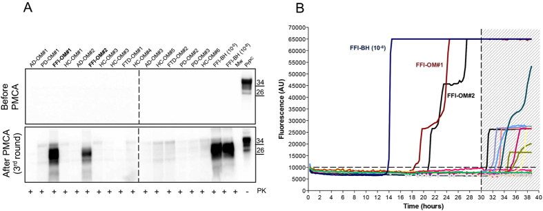
Figure 2. PrPSc detection in OM of FFI subjects using PMCA and RT-QuIC assays.
(A) Representative image of PMCA analysis of olfactory mucosa samples (n = 28) that were blindly performed. After three rounds of amplification PrPSc could be detected in two samples (FFI-OM#1 and FFI-OM#2) that belong to subjects that were symptomatic at the time of OM collection. 10−5 and 10−8 refer to dilutions of FFI brain homogenate in PMCA substrate that were used as positive control for reaction. The signal of PrPSc was assessed by means of Western blotting, after PK digestion, with the 6D11 antibody. Numbers in the right indicate the position of Mw. PrPC refers to normal bank vole brain homogenate that was not digested with PK. Dashed line indicates cropped images from separate gels. (B) RT-QuIC reactions were seeded with olfactory mucosa specimens of all patients. FFI brain homogenate (FFI-BH 10−8) was used as positive control for the reaction. Two OM samples were found positive before the threshold set at 30 hours. Average ThT fluorescence were plotted against time.

