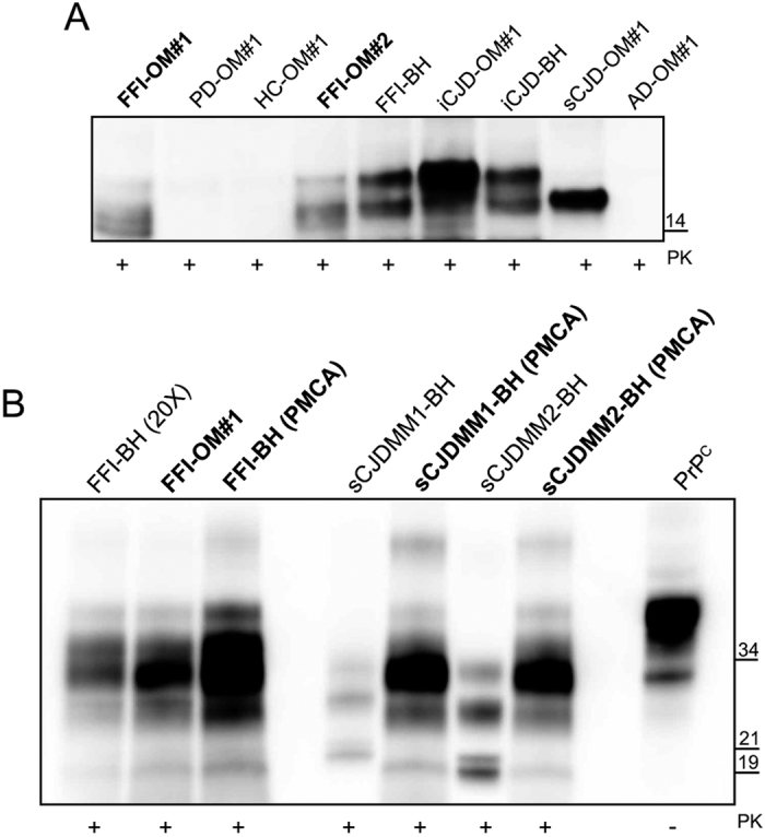
Figure 3. Amplified OM-PrPSc comparison with that of FFI or sCJD-type 1 and sCJD-type 2 brain homogenates.

(A) Western blot of the final products of RT-QuIC reaction seeded with brain homogenates or olfactory mucosa from sCJD, iCJD, FFI, AD, PD patients and healthy controls (HC). Samples were digested with PK [50 μg/mL] and immunoblot was probed with the C-terminal antibody SAF-84. Number in the right indicates the position of Mw. (B) Biochemical profile of amplified products (both brain homogenates and olfactory mucosa) was compared to that of FFI, sCJD-type 1 and sCJD-type 2 brain homogenates assessed before and after PMCA. FFI brain homogenate was concentrated 20-fold for PrPSc detection. The signal of PrPSc was assessed by means of Western blotting, after PK digestion, with the 6D11 antibody. Numbers in the right indicate the position of Mw: 21 kDa refers to type 1 PrPSc, while 19 kDa refers to type 2 PrPSc.
