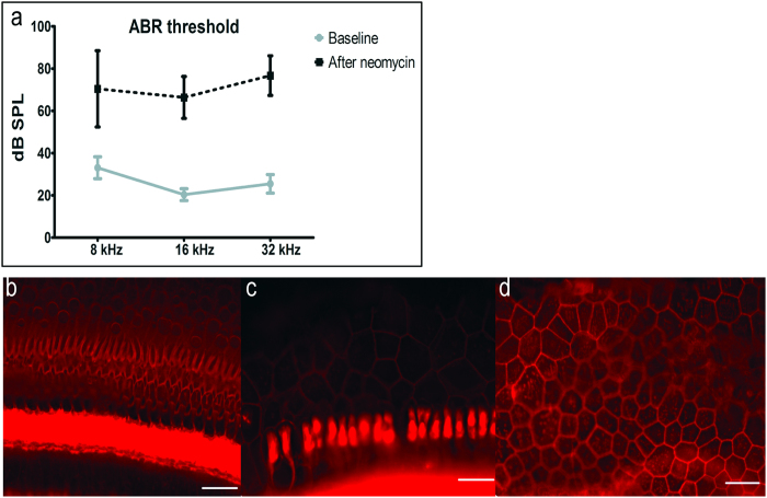
Figure 2. ABR thresholds and histologic changes after unilateral neomycin deafening.
(a) There was statistically significant elevation of thresholds overall (MANOVA, p < 0.01) and at each tested frequency (p < 0.05). Epi-fluorescence of auditory epithelium stained by phalloidin (actin, red) showed diverse tissue morphologies: scar formation (b), transition from scar formation to flat epithelium (c) or flat epithelium (d), but always complete hair cell loss. Scale bars are 25 μm.

