Figure 3.

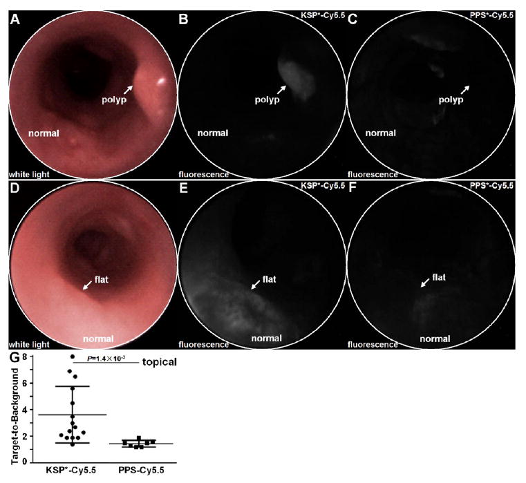
In vivo imaging of mouse colonic dysplasia with topical HER2 peptide. (A) Endoscopic image with white light illumination shows a spontaneous polyp (arrow). (B) NIR fluorescence image after topical administration of KSP*-Cy5.5 shows increased intensity from polyp (arrow). (C) Image of same region with PPS*-Cy5.5 control several days later shows significantly reduced signal. (D) White light and NIR fluorescence images with (E) KSP*-Cy5.5 and (F) PPS*-Cy5.5 show similar results with a flat adenoma (arrow). (G) From n = 7 mice, topically administered KSP*-Cy5.5 (n = 15 adenomas) resulted in a higher mean (±std) T/B ratio than PPS*-Cy5.5 (n = 7 adenomas), 3.64 ± 2.13 and 1.46 ± 0.25, respectively, P = 1.4 × 10⁻³ by unpaired t test.
