Figure 5.

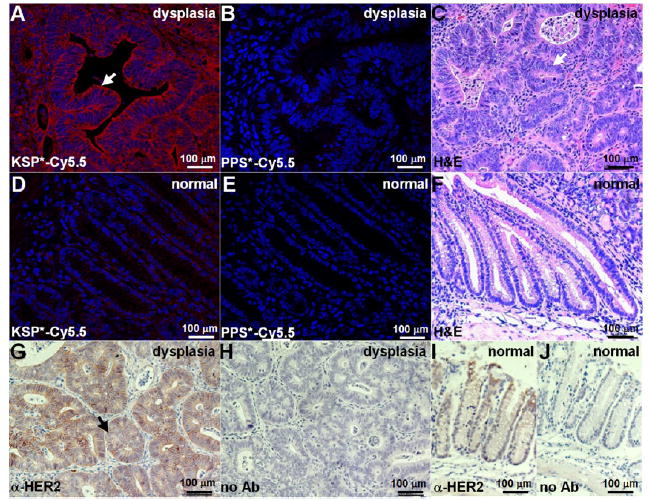
Validation of specific peptide binding to HER2 overexpressed by mouse colonic dysplasia. On confocal microscopy, we found intense staining of (A) KSP*-Cy5.5 compared to (B) PPS*-Cy5.5 to sections of dysplasia. (C) Histology (H&E) shows features of low-grade dysplasia (arrows). Minimal staining was observed with either (D) KSP*-Cy5.5 or (E) PPS*-Cy5.5 to normal colon. (F) Histology (H&E) for normal. (G) On immunohistochemistry with a known antibody, we confirmed overexpression of HER2 in dysplasia. (H) No antibody (control) with dysplasia. Normal colonic mucosa (I) with and (J) without antibody (control).
