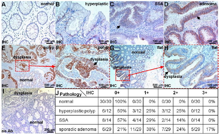
Figure 7.

Overexpression of HER2 in human proximal colonic neoplasia. On immunohistochemistry (IHC) of archived specimens, minimal staining was observed from all sections of (A) normal colon and most sections of (B) hyperplastic polyps. Intense cell surface staining was seen from some (C) sessile serrated adenomas (SSA) and (D) many sporadic adenomas. (E) Differences in HER2 expression between dysplastic and normal crypts from a polypoid adenoma can be seen. (F) Magnified view from dashed red box in (E) shows intense staining (arrow) from dysplastic colonocytes. (G) Difference in staining between dysplastic and normal crypts is shown for flat adenoma. (H) Magnified view from dashed red box in (G). (I) No antibody (control). (J) Consensus between 2 expert gastroenterology pathologists using a standard IHC scoring system revealed overexpression, defined by either 2+ or 3+ staining, in 0% (0/30) of normal, 25% (3/12) of hyperplastic polyps, 14% (2/14) of SSA, and 41% (12/29) of adenomas.
