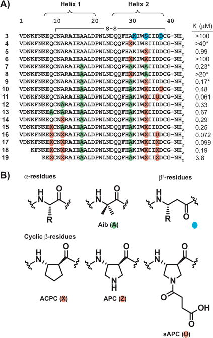
Figure 4.

A) Primary sequences of α/β-peptide derivatives of 2 and α-VEGF-2, along with associated Ki values, as determined by VEGF165 competition fluorescence polarization (FP) assay. Non-natural residues are indicated by colored circles. Each cysteine is engaged in an intramolecular disulfide bond. Ki values marked with a * are values previously reported for these compounds.[26] B) Structures of a generic α residue, the Aib residue (green), a generic β3 residue (teal), and the cyclic β residues ACPC, APC, and sAPC (orange). See Figure S1 for derivation of Ki values and a discussion of experimental uncertainty in FP measurements.
