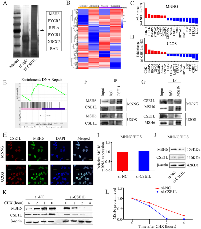
Figure 4. CSE1L interacts with MSH6 and affects its stability in osteosarcoma cells.
(A) Silver staining of SDS-PAGE gel after IgG or CSE1L pulldown. (B) Heat map of differentially expressed genes between the si-NC-treated group and the si-CSE1L-treated group in osteosarcoma cells. (C,D) mRNA expression levels of 7 upregulated and 10 downregulated genes selected for validation using qRT-PCR. (E) Gene Set Enrichment Analysis (GSEA) demonstrated enrichment of a gene signature associated with DNA repair. (F) Whole-cell lysates from osteosarcoma cell lines were immunoprecipitated with an anti-CSE1L antibody followed by immunoblotting (IB) with anti-MSH6 and anti-CSE1L antibodies. IgG was used as a negative control. (G) Whole-cell lysates from osteosarcoma cell lines were immunoprecipitated with an anti-MSH6 antibody followed by IB with anti-CSE1L and anti-MSH6 antibodies. IgG was used as a negative control. (H) Immunofluorescence analysis was performed using anti-CSE1L and anti-MSH6 antibodies. DAPI was used as a control for nuclear staining. (I,J) MNNG/HOS cells were transfected with si-NC or si-CSE1L for 48 h. The effect of CSE1L knockdown on MSH6 mRNA and protein expression in MNNG/HOS cells was evaluated by qRT-PCR and western blotting. β-actin was used as an internal control. (K,L) MNNG/HOS cells were transfected with si-NC or si-CSE1L for 48 h, followed by 0, 1, 2, and 4 h treatment with 100 mg/mL cycloheximide (CHX). Lysates were IB with an anti-MSH6 or anti-CSE1L antibody. MSH6 band intensity was normalized to β-actin, then normalized to controls (time = 0). For western blotting, full-length gels are presented in Supplementary Figure S10.

