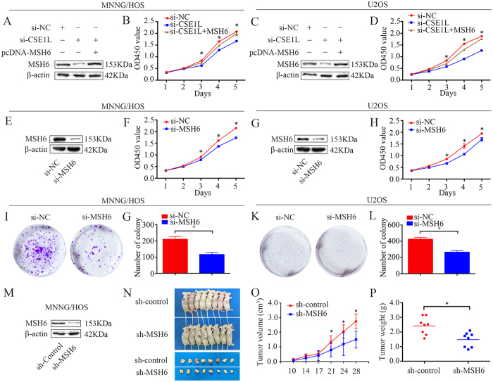
Figure 5. CSE1L knockdown inhibits osteosarcoma cell proliferation via MSH6.
(A,C) Representative blots of MSH6 after transfection with pcDNA 3.1-MSH6, si-CSE1L or si-NC. β-actin was used as an internal control. (B,D) Cell Counting Kit-8 (CCK-8) assay was used to measure tumor cell proliferation after transfection with pcDNA 3.1-MSH6, si-CSE1L or si-NC. (E,G) Confirmation of protein expression levels following si-MSH6 transfection by western blotting in MNNG/HOS and U2OS cells. (F) (H) CCK-8 assay was performed to detect tumor cell proliferation after siRNA transfection. (I–L) Colony formation assays for MSH6-silenced osteosarcoma cells and control cells. (M) Representative blots displaying CSE1L protein expression in MNNG/HOS cells stably expressing sh-control or sh-MSH6. β-actin was used as an internal control. (N–P) Representative tumor-bearing mice, tumors isolated from nude mice, tumor volume, and tumor weights of an MNNG/HOS subcutaneous tumor model. Data are representative of results from three independent experiments. *P < 0.05. For western blotting, full-length gels are presented in Supplementary Figure S11.

