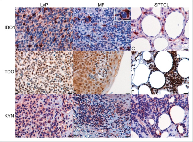
Figure 3.

Expression of IDO1, TDO, and KYN in tissue specimens of CTCL as determined by immunohistochemistry. (A) Expression of IDO1 in LyP. (B) Expression of IDO1 in TME of MF specimen and in macrophages surrounding the malignant cells (enlarged insert, upper right corner). (C) IDO-positive cells surrounding the adipocytes in SPTCL, (D) Expression of TDO in LyP. (E) Expression of TDO in MF. (F) Expression of TDO in SPTCL (G) Expression of KYN in LyP. (H) Expression of KYN in MF. (I) Expression of KYN in SPTCL. (Scale bar 20 µm and 40x magnification).
