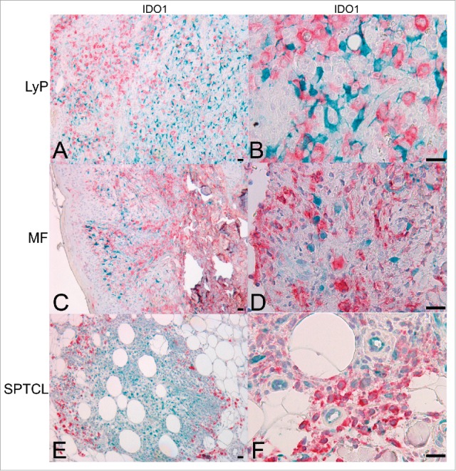
Figure 4.

IDO1 is expressed in CD163+ TAMs in SPTCL but not in MF or LyP skin lesions. Double IHC staining for IDO1 (turquoise) and CD163 (red) showing (A and B) LyP lesions and (C and D) MF lesions with separate cells expressing either IDO1 or CD163 (10x, and 40x, respectively), (E and F) Double positive IDO1+/CD163+ TAMs are seen surrounding the periadipocytic SPTCL infiltrate like a protective wall (10x and 40x, respectively). Light hematoxylin counterstaining. (Scale bar 20 µm). TAM, tumor-associated macrophage.
