Figure 5.

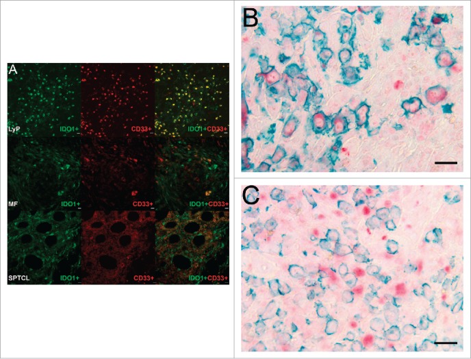
CD33+/IDO1+ expressing MDSCs are found in CTCL. (A) Double immunofluorescence staining of CD33 (red) and IDO1 (green) in typical LyP, MF, and SPTCL specimens. The top panel row shows a LyP tissue section with many strong double positive IDO1+/CD33+ cells (yellow) and some single positive IDO1+ or CD33+ cells. The second panel row shows an MF tissue section with cells either positive for IDO1+ or CD33+ as well as a few double positive IDO1+/CD33+ cells at lower right. The third panel row from top shows a SPTCL with double positive IDO1+/CD33+ cells as well as single positive cells (40x). In addition, IDO1 is expressed by both (B) CD30-positive and (C) CD30-negative cells in LyP (40x magnification). (Scale bar 20 µm).
